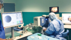
Retinal surgery
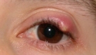
Retinal detachment is a disorder of the eye in which the retina separates from the layer underneath. Without treatment permanent loss of vision may occur
Signs and symptoms
- A sudden increase in size and number of floaters, indicating a retinal tear may be occurring;
- A sudden appearance of flashes, which could be the first stage of a retinal tear or detachment;
- Having a shadow appear in the periphery (side) of your field of vision;
- Seeing a gray curtain moving across your field of vision;
- A sudden decrease in your vision.
In the event of an appearance of sudden flashes of light or floaters, an eye doctor needs to be consulted immediately.
Risk factors
Risk factors for retinal detachment include severe myopia, retinal tears, trauma, family history, as well as complications from cataract surgery
Other risk factors include the following:
- Glaucoma
- AIDS
- Diabetic retinopathy
- Eclampsia
- Homocysteinuria
- Malignant hypertension
- Metastatic cancer, which spreads to the eye (eye cancer)
- Retinoblastoma
- Smoking
Diagnosis
Your ophthalmologist can diagnose retinal tear or retinal detachment during an eye examination where he or she dilates (widens) the pupils of your eyes. An ultrasound of the eye may also be performed to get additional detail of the retina.
Some retinal detachments are found during a routine eye examination. That is why it is so important to have regular eye exams.
Torn retina surgery
Laser surgery (photocoagulation)
With laser surgery, your ophthalmologist uses a laser to make small burns around the retinal tear. The scarring that results seals the retina to the underlying tissue, helping to prevent a retinal detachment.
Freezing treatment (cryopexy)
Your eye surgeon uses a special freezing probe to apply intense cold and freeze the retina around the retinal tear. The result is a scar that helps secure the retina to the eye wall
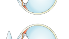
Detached retina surgery
Almost all patients with retinal detachments must have surgery to place the retina back in its proper position. Otherwise, the retina will lose the ability to function, possibly permanently, and blindness can result.
Scleral buckle
This treatment involves placing a flexible band (scleral buckle) around the eye to counteract the force pulling the retina out of place. The ophthalmologist often drains the fluid under the detached retina, allowing the retina to settle back into its normal position against the back wall of the eye. This procedure is performed in an operating room.
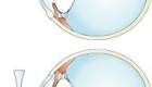
Pneumatic retinopexy
In this procedure, a gas bubble is injected into the vitreous space inside the eye in combination with laser surgery or cryotherapy. The gas bubble pushes the retinal tear into place against the back wall of the eye. The gas bubble will gradually disappear.
Vitrectomy
This surgery is performed in an operating room. The vitreous gel, is removed from the eye and usually replaced with a gas bubble.
Sometimes an oil bubble is used (instead of a gas bubble) to keep the retina in place. Your body’s own fluids will gradually replace a gas bubble. An oil bubble will need to be removed from the eye at a later date with another surgical procedure. Sometimes vitrectomy is combined with a scleral buckle.
Most retinal detachment surgeries (80 to 90 percent) are successful, although a second operation is sometimes needed.
Some retinal detachments cannot be fixed. The development of scar tissue is the usual reason that a retina is not able to be fixed. If the retina cannot be reattached, the eye will continue to lose sight and ultimately become blind.
After successful surgery for retinal detachment, vision may take many months to improve and, in some cases, may never return fully.
Information
-
 17 Jul 2023
 -
 03 Oct 2018
 -
 17 Apr 2018
 -
 13 Apr 2018
 -
 12 Apr 2018


© Copyright Vision-al.com All Rights Reserved.
