Corneal topography
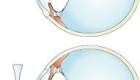
Corneal topography, also known as photokeratoscopy or videokeratography, is a non-invasive medical imaging technique for mapping the surface curvature of the cornea, the outer structure of the eye. Since the cornea is normally responsible for some 70% of the eye's refractive power, its topography is of critical importance in determining the quality of vision and corneal health.
The three-dimensional map is therefore a valuable aid to the examining ophthalmologist or optometrist and can assist in the diagnosis and treatment of a number of conditions; in planning cataract surgery and intraocular lens(IOL) implantation (plano or toric IOLs); in planning refractive surgery such as LASIK, and evaluating its results; or in assessing the fit of contact lenses. The procedure is carried out in seconds and is completely painless.
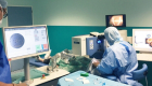
The patient is seated facing a bowl containing an illuminated pattern, most commonly a series of concentric rings. The pattern is focused on the anterior surface of the patient's cornea and reflected back to a digital camera at the centre of the bowl. The topology of the cornea is revealed by the shape taken by the reflected pattern. A computer provides the necessary analysis, typically determining the position and height of several thousand points across the cornea.
It is very important in the diagnose of keratoconus.
Information
-
 17 Jul 2023
 -
 03 Oct 2018
 -
 17 Apr 2018
 -
 13 Apr 2018
 -
 12 Apr 2018


© Copyright Vision-al.com All Rights Reserved.
