Strabismus
Strabismus is a visual problem in which the eyes are not aligned properly and point in different directions. One eye may look straight ahead, while the other eye turns inward, outward, upward, or downward. The eye turn may be consistent, or it may come and go. Which eye is straight (and which is misaligned) may switch or alternate.
Infantile esotropia, where the eye turns inward, is a common type of strabismus in infants. Young children with esotropia cannot use their eyes together. Accommodative esotropia is the most common form of esotropia that occurs in children usually 2 years or older. In this type of strabismus, when the child focuses the eyes to see clearly, the eyes turn inward. This crossing may occur when focusing at a distance, up close or both.
Exotropia, or an outward-turning eye, is another common type of strabismus. This occurs most often when a child is focusing on distant objects. The exotropia may occur only from time to time, particularly when a child is ill or tired. Parents often notice that the child squints one eye in bright sunlight.
Six eye muscles, controlling eye movement, are attached to the outside of each eye. . The brain controls these muscles.
To line up and focus both eyes on a single target, all of the muscles in each eye must be balanced and working together.
With normal vision, both eyes aim at the same spot. The brain then combines the two pictures into a single, three-dimensional image. This three-dimensional image gives us depth perception.
When one eye is out of alignment, two different pictures are sent to the brain. In a young child, the brain learns to ignore the image of the misaligned eye and sees only the image from the straight or better-seeing eye. The child then loses depth perception.
Adults who develop strabismus often have double vision because their brains have already learned to receive images from both eyes and cannot ignore the image from the turned eye. A child generally does not see double.
Good vision develops during childhood when both eyes have normal alignment. Strabismus may cause reduced vision, or amblyopia, in the misaligned eye.
Amblyopia can be treated by patching or blurring the stronger eye to strengthen and improve vision in the weaker eye. If amblyopia is detected in the first few years of life, treatment is usually successful. If treatment is delayed, amblyopia may become permanent. As a rule, the earlier amblyopia is treated, the better the result for vision.
Pseudostrabismus
The eyes of infants often appear to be crossed, though actually they are not. This condition is called pseudostrabismus. Young children often have a wide, flat nose and a fold of skin at the inner eyelid that can make eyes appear crossed. An ophthalmologist can distinguish true strabismus and pseudostrabismus.
Strabismus can be diagnosed during an eye exam. It is recommended that all children between 3 and 3½ years of age have their vision checked
If there is a family history of strabismus or amblyopia, or a family history of wearing thick eyeglasses, an ophthalmologist should check vision even earlier than age 3.
Treatment
Treatment for strabismus works to straighten the eyes and restore binocular (two-eyed) vision. In some cases of strabismus, eyeglasses can be prescribed for your child to straighten the eyes. Other treatments may involve surgery to correct the unbalanced eye muscles Patching or blurring the strong eye to improve amblyopia is often necessary.
Very young children with esotropia usually require surgery to realign the eyes.
For accommodative esotropia, glasses reduce the focusing effort and often straighten the eyes. Sometimes bifocals are needed for close work. If significant crossing of the eyes persists with the glasses, surgery may be required.
With exotropia, though glasses, exercises, patching or prisms may reduce or help control outward-turning of the eye in some children, surgery is often needed.
Strabismus surgery
In children with some types of constant strabismus, early surgery may be recommended to improve the chance of restoring or promoting normal binocular vision.
In adults, eye alignment surgery is not strictly cosmetic. There are many other benefits beyond restoring normal appearance: improved depth perception or binocular vision, improved visual fields, eliminating or minimizing double vision and improved social function.
It is important to discuss the goals and expectations of the surgery with your ophthalmologist
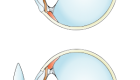
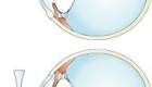
The ophthalmologist makes a small incision in the tissue covering the eye to reach the eye muscles.
One or more of the eye muscles are strengthened, weakened or moved to a different position to improve alignmentduring the surgery, depending on which direction the eye is turning. It may be necessary to perform surgery on one or both eyes.
Recovery time is rapid. Children are usually able to resume their normal activities within a few days.
After surgery, glasses may still be required. In some cases, more than one surgery may be needed to straighten the eyes.
Before surgery, a specialized examination called a sensorimotor examination will be performed in the Eye M.D.'s office to assess the alignment of the eyes to determine which muscles are contributing to the strabismus and which muscles need to be altered (weakened, strengthened, or moved) to improve the alignment of the eyes. Prisms are used to measure the degree of the strabismus. These preoperative tests help guide the surgeon in determining the surgical plan.
Strabismus surgery in children requires general anesthesia. In adults, the procedure can be done with general or local anesthesia.
Pain is minimal and usually over-the-counter medicines, such as ibuprofen (Motrin) or acetaminophen (Tylenol), and cool compresses are adequate.
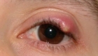
The eye will be red for one to two weeks, rarely longer, especially if it is a reoperation.
Potential risks of strabismus surgery
The chance of any serious complication from strabismus surgery that could affect the sight or well-being of the eye is exceedingly rare. However, there are risks with any surgery, including:
- Sore eyes;
- Redness;
- Residual misalignment;
- Double vision;
- Infection;
- Bleeding;
- Corneal abrasion;
- Decreased vision;
- Retinal detachment;
- Anesthesia-related complications.
Information
-
 17 Jul 2023
 -
 03 Oct 2018
 -
 17 Apr 2018
 -
 13 Apr 2018
 -
 12 Apr 2018


Contact Us
Follow Us
© Copyright Vision-al.com All Rights Reserved.
